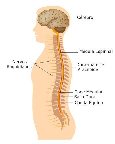
The spinal or spinal cord is a cylindrical cord, composed of nerve cells, located in the internal canal of the vertebrae.
Its function is to establish communication between the body and the nervous system, and also act on reflexes, protecting the body in emergency situations where there is a need for a quick response.
Despite being confused with bone marrow, it is related to the production of blood cells, while the spinal cord is part of the central nervous system.
Anatomy and Physiology
The spinal cord has a cylindrical shape, of non-uniform diameter, presenting two more dilated regions from which nerve fibers depart for the upper and lower limbs.
It consists of nervous tissue, located inside the spine and extends from the end of the brainstem (end part of the brain, comprises the midbrain, bridge and spinal bulb), after the bulb, to more or less the region of the second vertebra. low back.
The medulla becomes thinner at the end forming the medullary cone. Below the vertebra, surrounding the cone and a terminal filament, are the meninges (dural sac) and nerve roots of the last nerves that together form the cauda equina.
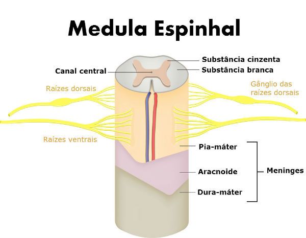
The inner region of the spinal cord, in the shape of an “H”, is called gray matter due to the high concentration of cell bodies of the neurons that give it this color.
While the outermost part contains more dendrites and axons and it becomes more whitish, being called white substance .
This arrangement of substances is contrary to that found in the brain. Externally, the medulla is surrounded by 3 membranes rich in collagen fibers, the meninges.
Meninges
These membranes contain spaces between them, which are lubricated by CSF or cerebrospinal fluid . CSF is a colorless, aqueous fluid that helps protect the central nervous system from impacts.
- Dura mater – more external and thick, it is like a sac that surrounds the whole medulla. It contains many blood vessels and is strongly adhered to the vertebral bones (and the skull, in the case of the brain). It has lateral extensions that involve the roots of the spinal nerves.
- Arachnoid – thin intermediate layer. It has delicate filaments that connect it to the pia mater, the arachnoid trabeculae, which give it an appearance similar to cobwebs.
- Pia mater – innermost, thin and delicate membrane. It is closely linked to the surface of the spinal cord (and brain). It confers resistance to the soft tissues of the nervous system.
Spinal Nerves
The nerves and nerve ganglia constitute the peripheral nervous system. Nerves are formed by branched nerve fibers that are distributed throughout the body and ganglia are dilations of some nerves where there is concentration of cell bodies of neurons.
Spinal or spinal nerves are mixed nerves because they contain sensitive and motor nerve fibers. They connect to the spinal cord in pairs, one on each side of the spine, through the spaces between the vertebrae.
Each nerve is composed of two sets of nerve fibers, called nerve roots, which connect to the dorsal part (dorsal root) and the ventral part (ventral root) of the spinal cord.
The dorsal root contains only sensitive nerve fibers, while the ventral root contains only motor nerve fibers.
At the dorsal root of each nerve is a ganglion made up of many cell bodies of sensory neurons.
Medullary Reflexes Acts
Reflex actions are quick, involuntary responses that are controlled by the gray matter in the marrow before it even reaches the brain, and are therefore important in defending the body in emergency situations.
For example, when we touch the hand in a very hot place, thanks to the reflex act we remove the hand immediately so as not to burn ourselves.
After receiving the stimulus, the sensitive fibers of the dorsal nerve root pass signals to associative neurons (located inside the medulla, in the gray matter), which in turn, pass them on to the motor fibers of the ventral nerve roots. These fibers send response to the organs that will carry out the action.