Inflammation of the synovial bag that is under the deltoid muscle, a muscle whose function is to lift the arm.
The subdeltoid and subacromial bursae lie below the upper portion of the deltoid muscle. They extend above and below the acromion and separate the greater tuberosity of the humerus from the deltoid muscle and the acromion. They eliminate friction and allow the greater tuberosity of the humerus to rotate inwardly, below the acromion, during abduction and rotation movements of the shoulder.
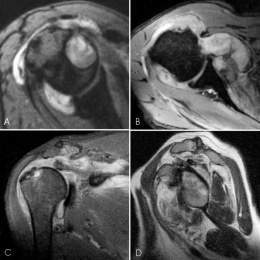
Subacromial bursitis is due to an adjacent injury that causes inflammation of this bursa; frequently this injury is that of the supraspinatus tendon.
Clinical manifestations.
Pain is characteristic of this condition and manifests as a tenderness referred to the upper portion of the shoulder, which radiates towards the insertion of the deltoid muscle.
The pain increases with abduction and internal rotation of the humerus, and is generally located at a point on the greater tuberosity, a point that disappears under the acromion when the shoulder is abducted (Dawbarn’s sign). In most cases there is a history of trauma in the days before the onset of pain.
Treatment
Conservative treatment. Treatment of acromial bursitis should be aimed at:
- Relieve pain, for which analgesics such as aspirin and dipyrone, among others, are used. Sometimes it is necessary to keep the arm at rest and for this an abduction splint is indicated.
- Remove inflammation, the purpose for which systemic anti-inflammatory drugs are used, such as cortisone and its derivatives, phenylbutazone and piroxicam. Local injection of 0.5% novocaine with hydrocortisone sometimes produces magnificent results.
- Apply physiotherapy; in this case, ultrasound combined with medication by means of phonophoresis helps in the definitive treatment of the condition, although sometimes, during the evolutionary phase, its application triggers acute painful crises.
- Treatment of large calcium masses when they persist despite conservative treatment; in this case it is necessary to extract them, and for this different methods are used:
- The bag is punctured with a No. 14 trocar, and then 1 ml of 0.5% novocaine and 4 ml of physiological saline are injected. Once the liquid has been introduced into the mouth, wait 5 min, mobilizing the shoulder; then the infiltrated liquid is aspirated and the calcium lumps are seen to come out in the aspirated liquid.
- The same previous procedure is performed but accompanied by multiple punctures with the trocar on the wall of the calcified bag, to loosen the calcium mass.
- The bag is washed, which seems to be the most efficient method. A No. 14 trocar is placed in the highest portion of the bag and another in the more sloping portion. 3 ml of 0.5% novocaine are injected, wait 5 min to produce anesthesia and then begin to wash the bag by dragging, introducing up to 200 ml of physiological saline through one trocar and letting it out through the other. The trocars are removed after leaving 2 ml of local hydrocortisone.
Surgical treatment
In extreme cases that do not respond to conservative methods, surgical removal is necessary. The operation consists of a vertical incision from the acromion on the deltiodes. It is penetrated through the muscle following the path of its fibers, the bag is reached and it is opened. Calcium deposits including those found in the supraspinatus tendon are removed. The wall of the bursa is carefully curetted and sutured flat. The arm is placed in an abduction airplane and the anti-inflammatory treatment is continued.