It is characterized by being in 90% of cases due to repeated compression, although in a small percentage they can be septic, staphylococcal and can be gouty. They are sometimes found in the course of rheumatoid arthritis or major post-trauma.
Other denominations
Cartoonist elbow, elbow student, elbow dart launcher, bursitis retroolecraneana, olecránica or olecranon, or elbow hygroma.
Causes
Olecranon bursitis can be:
1. Septic in nature, caused by various microorganisms, bacteria , viruses and fungi .
2. Aseptic. The triggering causes of aseptic bursitis are:
- Direct trauma to the bursa (contusions).
- Microtrauma, by repeated friction, as occurs in cartoonists, students, dart thrower.
3. Metabolic ( gout ).
Symptoms
In many cases, only the bag is found, not painful, bothering only because of its size and its unsightly appearance. In other cases there is mild pain or peribursal cellulitis or symptoms of systemic disease.
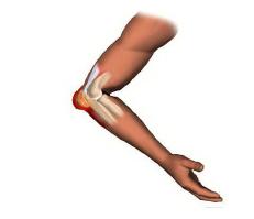
In a “dart thrower’s elbow” injury, a fluctuating oval mass forms at the tip of the elbow, painful, and swelling accompanied by redness of the skin that may be accompanied by subcutaneous bleeding .
Sometimes olecranon spurs and amorphous calcium deposits can be found (possibly due to the greater friction that this entails with the bursa) and in many cases these spurs are the same insertion of the triceps brachii. In the case of septic bursitis there is a marked increase in local temperature
Diagnosis
At the beginning the radiograph simply usually be negative. In chronic chaos, calcium deposits can appear . Ultrasound and MRI are helpful .
Puncture
If the liquid is clear it will be an aseptic bursitis. If the fluid is cloudy, an infection should be suspected , cultured, and empirical antibiotic treatment started that covers the possible microorganisms causing bursitis.
Treatment
1. Puncture of the bursa with needle 20, compression and rotation of the needle to extract the greatest amount of liquid, place the bevel of the needle towards the bone surface, without touching the bone .
Treatment with oral antibiotics according to antibiogram and parenterals is important if there is cellulitis.
2. Ice on the bursa (5 minutes for 2 days).
3. Compression bandage for 2 days or until pain and discomfort subsides.
4. Immobilization with neoprene sleeve or an elbow designed to offer comfort to the elbow joint.
5. Infiltration of lidocaine 0.5 cc plus corticosteroid 0.5 cc is the base of the bursa parallel to the ulna, with the usual penetration of 1 cc. Infiltration can be repeated a month. The joint should never be infiltrated if an infection is suspected .
The use of repeated corticosteroid injections can lead to infections, skin atrophies, and pain .