Bronchiolitis is the result of the inflammatory reaction produced after injury to the bronchiolar epithelium and the subsequent repair process, which leads to an excessive proliferation of granulation tissue. Depending on the intensity of the repair process, there is narrowing and distortion of the bronchioles (constrictive bronchiolitis) or their complete occlusion (obliterative bronchiolitis).
Obliterative bronchiolitis can have an idiopathic origin or present associated with different diseases and must be distinguished from bronchiolar inflammation that occurs in other diseases of the airway (COPD) or from those processes that occur with inflammatory changes in the lung parenchyma (obliterative bronchiolitis with organized pneumonia ).
Etiology
The clinical classification of obliterative bronchiolitis is based on the etiology of the process. Classically it has been recognized that obliterative bronchiolitis is a complication that occurs after inhalation of toxins or after viral infections of the respiratory tract. However, one of the most common forms of presentation today is that associated with bone marrow, lung and heart – lung transplants .
Obliterative bronchiolitis is one of the most common non-infectious complications after bone marrow transplantation, especially in cases of graft-versus-host disease. After lung or cardiopulmonary transplantation, the appearance of obliterative bronchiolitis is considered a form of rejection of the transplanted organ. Cases of obliterative bronchiolitis associated with connective tissue diseases, particularly rheumatoid arthritis , have also been described , in which it can appear as a complication of the disease itself or of treatment with penicillamine.
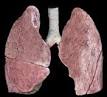
Pathological anatomy
On gross examination, the lungs appear distended, with lesions that are patchy. Microscopic examination reveals constrictive bronchiolitis with inflammatory infiltrate of the bronchiolar wall consisting of neutrophils and lymphocytes and concentric fibrosis of the submucosa. These lesions cause concentric narrowing of the lumen with bronchiolar obstruction. When it is incomplete, the distal alveoli are usually over-distended, while when it is complete, they appear atelectatic and fibrotic.
The most distal segments of the airway, the alveolar spaces and the pulmonary interstitium are preserved and have normal characteristics. These histopathological features are distinguished from those of proliferative-type bronchiolitis (obliterative bronchiolitis with organized pneumonia ), in which the formation of intraluminal granulation tissue that occupies the respiratory bronchioles and alveolar ducts is characteristic, and is usually associated with interstitial inflammatory infiltrate (see Diffuse Interstitial Diseases of the Lung ).
Clinical picture
The most common symptoms are non-productive cough and dyspnea on exertion. In cases that occur after inhalation of toxins or viral infections, fever and chest pain may be associated. Unlike other obstructive airway diseases, obliterative bronchiolitis has a relatively short onset, with a few months of evolution. Physical examination may be normal or characteristic of bronchial obstruction, auscultating long expiration, rhonchi, and wheezing. In some cases, the presence of diffuse inspiratory rales has also been described. On chest x- ray the most characteristic finding is pulmonary insufflation, which in some cases may be accompanied by small diffuse opacities and reinforcement of the bronchovascular fabric, indicating the existence of focal areas of atelectasis and peribronchiolar infiltrate.
The respiratory function examination reveals an obstructive ventilatory alteration, with a reduction in FEV1 and the FEV1 / FVC ratio, which does not usually revert after the administration of a bronchodilator. The FVC can also be reduced by the presence of air trapping and, on occasions, by a restrictive ventilatory alteration due to the presence of areas of atelectasis. In bronchoalveolar lavage, intense neutrophilia is observed, usually greater than 50% of the cells obtained.
Diagnosis
The accurate diagnosis is established by lung biopsy. However, when a biopsy involves a high risk for the patient, the diagnosis can be established if there is: a) a clear clinical history (transplantation, inhalation of toxins); b) rapid onset clinical course; c) airflow obstruction that is not due to other diseases and that does not respond to the administration of bronchodilators, and d) frank neutrophilia in bronchoalveolar lavage.
The differential diagnosis should be made especially with other obstructive diseases of the airways. As has been pointed out, obliterative bronchiolitis is a different disease from obliterative bronchiolitis with organized pneumonia , both due to the pathological characteristics and the clinical picture. Unlike obliterative bronchiolitis, obliterative bronchiolitis with organized pneumonia is an interstitial disease characterized by the presence of alveolar infiltrates on the chest X- ray , a restrictive respiratory function alteration and a good response to glucocorticoid treatment. .
Treatment
Treatment of obliterative bronchiolitis aims to reduce bronchiolar inflammation through the administration of glucocorticoids. Prednisone is administered at a dose of 1 mg / kg / day for about 3 months, and then it is progressively reduced to a daily dose of 20 mg, which is maintained for a period of 1 or 2 years. The response to this treatment is only favorable in half of the cases.
Although the immediate bronchodilator response is usually negative, the administration of bronchodilators may be beneficial in some patients. The treatment of obliterative bronchiolitis associated with lung or cardiopulmonary transplantation consists of the administration of glucocorticoid boluses and increased immunosuppressive treatment, since this process constitutes a manifestation of rejection of the transplanted organ. Glucocorticoids, bronchodilators, and immunosuppressive drugs have been used for the treatment of obliterative bronchiolitis associated with bone marrow transplantation, generally with unsatisfactory results. For this reason, early detection of this complication is important, based on a high index of clinical suspicion and performing respiratory function tests.