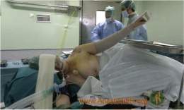
The appearance of bronchospasm during the administration of anesthesia is not a frequent complication, but it is a situation that alarms and turns into an unpleasant experience that can cause nocturnal dyspnea , a feeling of congestion and tightening of the chest that makes them wake up, as well as congestion nasal and respiratory distress variable to irritants, such as cold air, have bronchial hyperresponsiveness , among others.

Appearance of the disease
Preoperative identification of patients at risk of developing bronchospasm during anesthesia allows a series of preventive measures to be taken.
This review is carried out with the objective of updating anesthesiologists on the pathophysiological and pharmacological mechanisms necessary to allow them to reduce the incidence of this complication and, when it occurs, be properly prepared to face it.
Diseases that cause hyperresponsiveness of the airways
- Bronchial asthma .
- Chronic bronchitis and emphysema .
- Allergic rhinitis .
- Upper and lower respiratory disease .
- Smokers with airway obstruction .
When talking about hyperresponsiveness of the airway associated with bronchospasm, one immediately thinks of asthmatic patients and those suffering from chronic bronchitis and emphysema. However, patients with infectious respiratory tract diseases and smokers are much more likely to develop this complication during anesthesia .
An extremely important trigger factor that is sometimes underestimated is a history of recent upper respiratory tract disease. The presence of a viral disease of the upper respiratory tract causes a marked hypersensitivity and reactivity of the bronchial tree, which persists for 3 to 4 weeks, even in the patient without a history of respiratory disease.
Asthmatics and bronchitis patients deteriorate significantly when they suffer from respiratory diseases, and they are forced to maintain a sustained bronchodilator treatment . Bronchial reactivity increases considerably in the presence of mucosal inflammation .
Previously asymptomatic patients who begin to suffer from respiratory symptoms such as nocturnal dyspnea , a feeling of congestion and tightness of the chest that makes them wake up, as well as nasal congestion and respiratory distress variable to irritants, such as cold air, have bronchial hyperresponsiveness and should be studied and treated preventively before undergoing any anesthesia.
Pre-operative treatment
- Agonists b- adrenoceptor .
- Aminophylline .
- Corticosteroids .
- Anticholinergics .
- Do not smoke or inhale irritants.
As a first measure, patients with respiratory disease should not undergo elective anesthesia. It is known that even after a “simple” viral upper respiratory infection, even normal people have an increased reactivity of the airways and will present complications with the administration of anesthesia.
The b-adrenergic agonist drugs are excellent bronchodilators and extremely useful in the treatment of bronchospasm during anesthesia.
This group of drugs, currently used b2 stimulants ( albuterol , terbutaline , salbutamol ) are safe and highly effective. After its administration, relaxation of the bronchial muscle is observed, with a low incidence of undesirable effects such as cardiac stimulation and tachyarrhythmias . It is preferable to use these drugs by inhalation, otherwise higher doses are needed and high plasma concentrations and a higher incidence of complications occur.
The aminophylline has a narrow margin of safety and can trigger cardiac arrhythmias during anesthesia, so its current use is reserved for the prevention of acute attacks in asthmatic patients and as a second – line drug in the treatment of bronchospasm, administered slowly intravenously. The administration of this drug produces an increase in endogenous catecholamines that is proportional to the dose.
In children with congenital heart disease undergoing surgical reconstruction under extracorporeal circulation (ECC) , it is used at a dose of 5 mg per kg of weight, by releasing the aortic root clamp, with the aim of improving respiratory function as this drug improves ciliary activity and diaphragmatic contraction , thus contributing to better ventilatory dynamics after CPB.
Parenteral corticosteroids are of great value in the prevention and treatment of intraoperative bronchospasm. They must be administered preoperatively or when the first vein is channeled, since it takes some time (60 to 90 min) to obtain their maximum therapeutic effects.
The Atropine is used to prevent bronchoconstriction reflects that may occur during handling of the airway and intubation of the trachea.
In patients with smoking habit, abstinence is accompanied by evident improvement, which is characterized by a significant decrease in the amount of bronchial secretions, less bronchial reactivity, and a better function of mucociliary transport activity.
Selection of anesthetic technique and agents
The regional anesthesia is the technique of choice for patients with airway hyperresponsiveness, as it eliminates the trigger that is the manipulation of the airway.
Intravenous induction agents
The thiopental itself produces no bronchospasm, but nevertheless manipulation airway during a light anesthesia with this agent triggers important reflexes producing laringo and bronchospasm.
Some authors suggest that ketamine produces relaxation of the bronchial smooth muscle by direct action, by releasing catecholamines and by decreasing the vagal response produced to stimuli, and they consider it an agent of choice in patients with respiratory disease.
The propofol is an agent useful in the induction of anesthesia in the patient at risk of bronchospasm. This agent profoundly depresses reflexes and a significant decrease in airway resistance is observed after administration.
The fentanyl produces histamine release, but can cause the stiffness of the chest and ventilatory significant difficulty. This effect is blocked with adequate muscle relaxation. This opioid can cause bronchoconstriction and increased pulmonary resistance by vagal stimulation, which causes contraction of the bronchial smooth muscle. Administration of atropine counteracts this effect.
Halogenated Agents
The halogenated agents such as halothane , enflurane and isoflurane produce broncho-dilation direct relaxing effect on the bronchial muscle and inhibition of airway reflexes. These agents have been considered over time, as those of choice in asthmatics. One limitation for their use is the cardiovascular depression they produce, which is why it is suggested to use them in low concentrations (halothane less than 1%) associated with other bronchondilators.
Local anesthetics
The lidocaine used as a preventive treatment to block the airway reflexes, in patients with bronchial hyperreactivity and for the treatment of bronchospasm intraoperatively. This agent prevents bronchospasm by blocking airway reflexes and by direct action on bronchial smooth muscle, and attenuates the response to acetylcholine .
The parasympathetic nervous system controls the basal tone and the changes produced in the bronchial muscle produced by the stimulation of the airway. Receptors within the wall of the airways change the tone of the bronchial muscle through vagal transmission pathways. Among the most important receptors are those found in the mucosa of the cartilaginous airways and especially in the trachea and carina. These receptors respond vigorously to stimuli such as irritation, temperature changes, particles, or inhaled irritating gases. Airway edema and histamine also increase their activity, causing coughing, mucous discharge, and bronchoconstriction. The efferent reflex travels through the parasympathetic and vagus pathways to synapse within the wall of the airways.
Lidocaine administered intravenously blocks this reflex and stimulates broncho-dilation. Atomization of the airways with lidocaine can cause irritation and bronchospasm, so its intravenous administration is preferred.
Diagnosis and treatment of bronchospasm
The patient who develops bronchospasm during anesthesia shows the following symptoms and signs:
Cough.
- Increase in insufflation pressure.
- Distended chest and decreased lung compliance.
- Air entrapment.
- Wheezing.
Increased secretions and mucosal edema contribute to complicating this situation. Some patients also develop air trapping with a further worsening of the clinical picture.
The measures used in the treatment of bronchospasm during anesthesia are as follows: [14]
- Increased depth of anesthesia . The use of high concentrations of halogenated agents is not a prudent measure, due to the hemodynamic deterioration that these agents produce.
- Administration of bronchodilators. The b2 stimulant drugs are very effective and safe agents and can be used in frequent doses by direct spray.
Give a large dose of muscle relaxant. In the anesthetized patient, it is necessary to produce deep muscle relaxation to eliminate coughing and muscle contraction that worsens ventilation.
- Intubation of the trachea and couple the patient to a mechanical ventilator. A powerful ventilator such as the Servo 900 D is needed to overcome the airway resistance that occurs frequently during this complication. Allow adequate expiration time.
Aminophylline (2 to 5 mg / kg) given slowly intravenously.
- Corticosteroids Administering ketamine intravenously at an average dose of 2 to 4 mg / kg is a quick and easy way to increase the depth of anesthesia while maintaining stable blood pressure.
