It is defined as a localized collection of pus in the liver , resulting from any infectious process with destruction of the liver parenchyma and stroma , being in order of frequency: BACTERIAL (pyogenic), MYCOTICS ( Candida Albicans ), which shows an increasing incidence parallel to the AIDS and that of bone marrow transplantation , chemotherapy and radiotherapy . AMEBIAN ( Entamoeba Histolítica ), as a rare complication of intestinal amebiasis .
Pyogenic abscess
It is due to a polymicrobial infection by gram-negative aerobic and gram-positive anaerobic germs, most often Escherichia coli in two-thirds, followed by S. faecalis, Klebsiella and Proteus bulgaris . Staphylococci, especially in patients who have received chemotherapy. The age of presentation ranges between 40 and 60 years, with a greater predilection for the female sex.
Pathways of dissemination
- BILE: (40%) due to superinfection, secondary to an obstacle in the drainage of the bile that favors intraparenchymal bacterial diffusion with the formation of multiple abscesses, affecting both lobes in 90% of cases.
- PORTAL: (25%) secondary to an intra-abdominal or poorly treated septic focus (sigmoiditis, ulcerative colitis, Crohn’s disease, superinfected cancers, pancreatitis). There is often thrombosis of a portal venous afference in contact with the primary infectious focus that causes bacteremia and liver seeding, they are usually unique and 65% affect the right lobe, 12% the left and 23% both. This distribution has been attributed to the mesenteric blood flow pattern of the portal vein.
- IDIOPATHIC: (20%) in diabetic patients, cryptogenic abscesses of unidentifiable origin may appear.
- BY CONTIGUITY: (25%) post-traumatic or post-surgical.
Clinical manifestations
In the pre-antibiotic era, the picture consisted of fever peaks and pain in the right upper quadrant with prostration and shock.
Currently, the presentation is less acute, with dull pain that increases with movement, malaise, hepatomegaly, low-grade fever, and weight loss. It is quite possible that it is hidden in the old man.
Pathological anatomy
The enlarged liver may contain multiple yellow abscesses 1 cm in diameter or a single abscess surrounded by a fibrous capsule. When there is pylephlebitis, the aorta vein and its branches may contain pus and blood clots, there may be perihepatitis with adhesion formation.
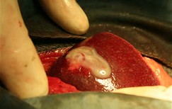
A single chronic liver abscess can exist up to two years before death and diagnosis.
Causes
There are many possible causes of liver abscesses, including:
- Abdominal infection, such as appendicitis, diverticulitis, or a perforated intestine
- Infection in the blood
- Bile duct infection
- Recent endoscopy of the bile ducts
- Trauma that damages the liver
Some of the more common bacteria that cause a purulent liver abscess include: 5
- Streptococcus milleri
- Escherichia coli
- Streptococcus faecalis
- Klebsiella
- Proteus vulgaris
- Opportunistic pathogens such as staph.
Laboratory findings
There is leukocytosis with an increase in PMN; Increased serum alkaline phosphatase; Very high ESR; Positive CRP, hypoalbulinemia and prolonged prothrombin time.
X-ray of abdomen
Hepatomegaly, with right diaphragmatic hump and pleural effusion. There may be a collection of gas bubbles on the liver (Klebsiella germs, amebiasis).
Ultrasound
The echostructure depends on the evolutionary stage of the abscess. The PRESUPURATIVE STAGE in which there is still no collection, there are poorly defined heterogeneous hyperechogenic or hypoechoic zones that take on the appearance of a usual COLLECTION composed of a hypoechoic central zone that sometimes contains internal echoes surrounded by a peripheral hyperechoic envelope of variable thickness with back reinforcement. The lesion can also simulate a homogeneous or heterogeneous solid mass.
The visualization of small hyperechogenic areas with cones of posterior shadow suggests microbubbles of gas produced by anaerobic germs.
In abscesses of fungal origin, they are generally multiple up to 2 cm in diameter with the characteristic of being hyperechoic with a hyperechoic center in “bull’s eye” or “cockade” sign.
THE DIFFERENTIAL DIAGNOSIS: With these images, you must include, hematoma, metastasis, necrotic tumor, hemangioma, hemorrhagic cysts and complicated hydatid cyst.
Computed tomography
It is the examination that provides the most information, being performed without and with contrast and its detection being greater than 90%. The attenuation limits range from 0 to 45 HU. In the PRESUPURATIVE STAGE, the lesions are hypodense and heterogeneous, not enhancing after contrast. In the COLLECTION STAGE, the hypodense lesion is surrounded by a more or less thick envelope that is clearly enhanced.
The lesions can cause disorders of the perfusion of the adjacent parenchyma in the form of hyperdense systematized areas in the arterial phase. The presence of gas bubbles or air-fluid levels in the absence of a biliodigestive fistula is pathognomonic for superinfection by anaerobes but is only found in less than 20%.
Magnetic resonance
In pyogenic abscesses, it has a low signal intensity on T1 and high signal on T2. With three important findings:
- In one third of cases, there is edema around the abscess in T2 that is absent in hemangiomas and solid masses.
- After contrast administration, there is a ring enhancement with a center that is generally hypointense or heterogeneous, this represents the fibrous capsule with inflammatory cells.
- They tend to have a “cluster” configuration like “a clover leaf.” This “cluster sign” is rarely identified in multifocal metastases.
Fungal microabscesses have clinical characteristics and, on MRI, the characteristics are usually lesions smaller than one centimeter associated with lesions in the spleen and / or kidney showing high intensity on T2 and enhancement after contrast.
Treatment
Establishing the cause of the abscess is of vital importance with respect to the last chosen therapeutic scope. However, we must take into account the INFLAMMATORY PSEUDOTUMOR OF THE LIVER, which is a rare benign process, characterized by fibrovascular proliferation with inflammatory cells (44 cases reported in the literature until 1995). Its etiology is unknown, with clinical and laboratory behavior such as an acute abscess, with the histopathological study confirming the diagnosis.
In addition to adequate antibiotic therapy, most require needle or catheter drainage and treatment of the obstruction if present. Amoebic and fungal abscesses are treated with Metronidazole and Amphotericin B respectively, and drainage is usually not required.
Currently, the radiologist is part of the multidisciplinary team in charge of diagnosis and treatment. As the images obtained by CT, US and MRI are often not specific for abscess, aspiration puncture is essential to confirm the diagnosis and start antibiotic treatment, in addition to being therapeutic in order to extract the maximum possible amount of pus to retract the capsule and promote better penetration of antibiotics. Systematically, a capsule biopsy should be performed when aspiration is negative in order not to ignore a necrotic tumor lesion or a granulomatous lesion. Percutaneous catheter drainage is more favorable in abscesses larger than 4 cm.
Prognosis and complications
Complications include septic shock, infection secondary to other organs, perforation with peritonitis, and fistula formation.
Needle aspiration and antibiotic therapy have reduced mortality to 16%. The prognosis is better in uniloculated abscesses of the right lobe with a survival of 90%. Survival for multiple abscesses, especially if they are biliary, is very poor and they only reach a survival of 20%. Prognosis worsens with delay in diagnosis, continued fever, blood culture-demonstrated polymicrobial infections, hyperbilirubinemia, associated diseases, hypoalbulinemia, pleural effusion, and advanced age.