Glial cells make up nerve tissue along with neurons. Glial cells, also called gliocytes or neuroglia, can be of two types: microglia or macroglia.
In addition to providing nutrients, protection and helping support nervous tissue, they have other important functions, such as the modulation of electrical impulses.
Occupation
Although they are much more numerous, constituting about 80% of the nervous tissue, for a long time they were considered only responsible for feeding neurons.
However, more recent studies have shown that gliae, in addition to nourishing, protecting and helping support nervous tissue, also regulate synapses by neurotransmitters. .
They are also responsible for neurogenesis , that is, the formation of new neurons.
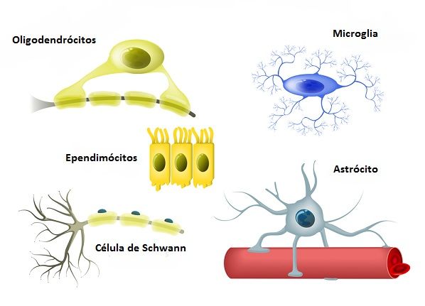
Microglia
They are much smaller cells than the other glia. They have a cell body with few extensions with some short ramifications. Microglia have a similar function to that of macrophages (cells of the immune system), that is, they make phagocytosis.
It is related to the protection of the nervous system. They are activated when there are lesions, infections or degenerative diseases, which causes it to proliferate intensely and perform the phagocytosis of invading agents such as viruses.
Macroglias
There are four types of macroglia most well-known: astrocytes, oligodendrocytes and Schwann cells.
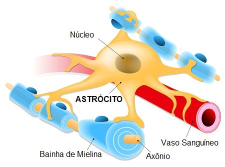
Astrocytes
Astrocytes are the largest and most common glial cells, making up about half of the brain. There are several subtypes related to different functions, especially the metabolism of neurotransmitters, their uptake and the functioning of synapses.
These cells make up the blood-brain barrier, which is a protection of the central nervous system to toxic agents present in the blood.
Schwann cells
Schwann cells are responsible for the formation of the myelin sheath in neurons in the peripheral nervous system. They wrap around the axons, electrically isolating them. The spaces between the cells form discontinuities in the myelin sheath forming Ranvier’s nodules.
Axon myelination makes the propagation of the electrical impulse faster and more efficient, which is also due to the jumps produced by the discontinuity of the nodules.
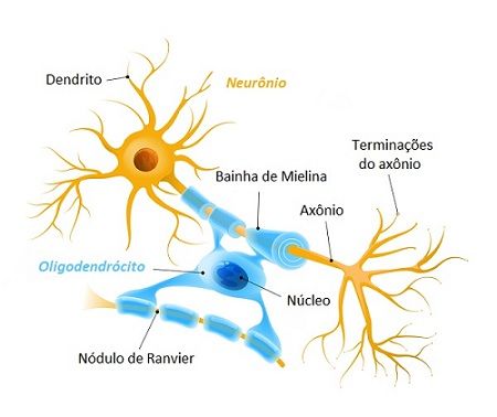
Oligodendrocytes
Oligodendrocytes have few extensions, so they have that name (oligo = little). They participate in the process of myelination of neurons in the central nervous system, that is, in the formation of the myelin sheath that surrounds and protects axons.
Ependymal Cells
Ependymal cells or ependymocytes are cells lining the nervous system. They line the ventricles of the brain and the central canal of the spinal cord.