Ulcers that appear on any mucous surface of the mouth. Its most frequent locations are, in descending order, the oral and labial mucosa, the edges of the tongue , the buccal and lingual grooves and the soft palate, but not on the gingiva.
Characteristics of Canker Sores
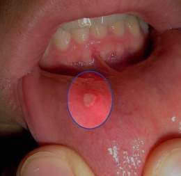
They are shallow, painful sores that appear inside your mouth. They are usually red in color or may sometimes have a white coating covering them. They can hit the inside of your lips, the inside of your cheeks, the base of your gums, or under your tongue. Canker sores are different from the sores (cold sores) that are usually on the outside of your lips or at the ends of your mouth.
Anyone can get canker sores, but women in their teens and second decade of life get them more often. Canker sores can be passed between family members but are not contagious. Doctors don’t know what causes canker sores, but they can be triggered by stress, poor nutrition, food allergies, and menstrual periods.
Fundamental manifestations.
They usually appear temporarily and physically isolated, although they can be multiple, fuse and cause a single lesion. Intense burning or itching usually occurs, which precedes the appearance of the ulcer. This can be unique or form groups of up to 30 or more. Sometimes, several canker sores can coalesce, forming lesions of irregular contour. They persist for about two weeks and subsequently disappear without scarring. Canker sores can be very painful and can affect a person’s daily activities. There is usually no fever or other symptoms, unless secondary infections occur. They tend to reappear with some frequency.
The injury begins with a burning sensation; then a small reddish macula appears, followed by a vesicle, which ruptures and the ulcer is formed consisting of a light yellow spot, generally oval, one millimeter to one centimeter in diameter. It can present with one or more lesions covered with a yellowish layer on a red base, which tend to recur. They usually start with a burning sensation at the site of the future ulcer. After several days they progress to a swelling that becomes ulcerative. The gray, white, or yellowish area is due to the formation of fibrin, a protein associated with blood clotting.
Location.
They occur on the lip mucosa, mucosal folds, on the floor of the mouth or on the gums, and can be solitary or multiple. The lesions are very painful and when they are multiple they can lead to inflammation of the cervical nodes, general malaise and fever. In general they are very sensitive to food and the pain can spread to the entire face, also causing difficulties in eating.
Etiology
In most cases the cause is unknown. There is talk of allergic and irritant factors such as drugs, food. They can also be related to bacterial and viral forms, and some autoimmune elements may even participate.
It is postulated that the following factors are involved:
- Genetic components.
- Immunological factors.
- Infectious components (viral or bacterial).
Other factors or conditions that can lead to the appearance of canker sores are:
- Erosive oral injuries and repetitive trauma affecting the oral mucosa.
- Hypersensitivity to certain foods, preservatives or food additives.
- Adverse reactions to certain medications.
- Stress and emotional disorders.
- Hormonal alterations
- Nutritional deficiencies ( iron , zinc , vitamin B12 and folic acid .
- Digestive disorders or manifestations of other diseases such as ulcerative colitis, Crohn’s disease, celiac disease, Behçet’s disease, Candidiasis , immunodeficiencies, etc.
Disorders.
The presence of oral sepsis and dental caries are related to the disease in many cases.Image of injuries.
Diagnosis.
The diagnosis is clinical. It is important to know the personal and family pathological antecedents of the patient ( diabetic , immunosuppressed , hematological disease, Crohn’s disease , adiopathic ulcerative colitis, etc.), oral trauma, use of prostheses or dental material, burns and infections, all obtained during the anamnesis and reflected in the medical history.Image of injuries.
At present, oral mucosa culture studies, biopsies , exfoliative cytology, viral detection of monoclonal antibodies or by PCR techniques, histological visualization of intracellular viral inclusions and serological studies can be carried out, in those cases that require it or are immunocompromised. .
Hygienic measures
Maintain oral hygiene, attend the stomatologist regularly and avoid possible causal factors such as medicines and foods that are known to trigger the disease. Fundamentally the following sanitary measures:
- Avoid or minimize possible triggers for canker sores.
- Use soft bristle toothbrushes, handle other dental hygiene accessories (dental floss, interdental brushes, etc.) gently.
- Brush your teeth after every meal.
- Make regular visits to the dentist.
- Change the toothbrush frequently (maximum every 3 months) and keep it in correct hygienic conditions.
epidemiology
The prevalence of canker sores is approximately 20%, with a higher incidence in children and adult women. Being rare in adults over 50 years of age.
Classification
According to their clinical manifestations, shapes and size, three types of canker sores can be clearly differentiated:
1. Minor Canker Sores .- They are small ulcers that are usually located in non-keratinized areas of the oral mucosa. In the same episode, between 1 and 5 mild lesions may appear and whose size does not exceed 10 mm in diameter. It does not usually have recurrences.
2. Major Canker Sores . – They can appear alone or several together, especially on the lips, tongue , pharynx , palate and internal area of the cheeks. Their size is greater than 10 mm in diameter and they are deeper and more destructive than the former. They usually leave a scar and hypochromic mucosa with superficial fibrosis.
3. Recurrent Aphthous Stomatitis .- In this case, multiple canker sores appear in groups and regularly in any part of the mouth . The size of the lesions does not usually exceed 3 mm, but they tend to coalesce to form larger ulcers.
Treatment.
Given the diversity of the etiology and knowledge of the immunological disorders present in RAS, the proposed therapeutic approach is aimed at modulating the inflammatory response, alleviating symptoms and avoiding recurrences. To this end, local and systemic treatments are used.
Medicines used for its treatment.
Currently:
- Schostacovsky balm .
- use of corticosteroids , hydrocortisone ).
- antihistamines (benadryline).
- antibiotics ( tetracycline ).
- Amlexanox .
- Although the most recommended by specialists is Nystatin in suspension.
Local treatment.
Analgesic-anti-inflammatory:
- Acetylsalicylic acid (ASA): little accepted as it is a chemical agent that causes lesions of the oral mucosa. It is used in oral rinses 15 minutes before meals.
- 5-aminosalicytic acid (5-ASA or mesalazine): it is applied in the form of a cream, 3 times a day after meals; relieves pain and shortens healing time.
Formula: 5-ASA 5% Cream o / w qs 10%
- Benzydamine: anti-inflammatory in the form of a spray or 0.1% mouthwash. Relieves pain and reduces ulcerated area.
Local anesthetics:
- Solutions or gel: xylocaine and 2% lidocaine. They are applied in the form of a gel, as viscous solutions or in rinses before meals, fast acting and relieves pain for an hour. It is recommended not to ingest to avoid systematic toxicity; You can add 1/100 000 adrenaline.
Formula: Lidocaine 2% Sodium carboxymethylcellulose 2% Aqueous solution qs 100 mL
- In major canker sores, intralesional injection of 2% lidocaine can be applied.
Antibiotics:
- Tetracyclines and derivatives (chlortetracyclines, doxycycline, minocycline): used as mouthwashes to reduce pain, prevent superinfection, and accelerate healing.
- Tetracycline (250 mg) 4 tablets dissolved in 180 mL of water, in rinses for 3 minutes every 4-6 h for 3-5 days.
Formula: as long as there is no suspicion of bacterial superinfection, the use of a simple antibiotic (formula 1) is recommended, but the antibiotic with antifungal and corticosteroids (formula 2) can also be applied.
Formula 1: Tetracycline 2.5-5% Nystatin 5-10 106 U Glycerin qs 50 gImage of injuries.
Formula 2: Tetracycline 2.5-5% Clotrimazole 1% Triamcinolone acetonide 0.1% Glycerin qs 50 g
- Cephalosporins: cephalexin (250 mg): dissolve one tablet in 30 mL of water and apply it for 10-20 minutes, every 4-6 hours.
Antiseptics:
- Chlorhexidine gluconate 0.12%, hexetidine 0.1%, hydrogen peroxide or hydrogen peroxide 1.5%, glycerated solutions of sodium borate and povidone iodine. It is used in slightly concentrated solutions applied with cotton swabs for 2 minutes every 8 hours, with a prophylactic purpose on mild bacterial and fungicidal superinfection.
Formula: Sodium borate 4 g Glycerin 30 g Antifungal
- Nystatin, clotrimazole: it is applied in mouthwashes or in formulas where they are combined with antibiotics.
Cytoprotectors:
- Sucralfate 1 g (urbal): it is a mucosal protector that forms a layer over the crater of the ulcer, which prevents irritants from acting on the lesion, allowing pain relief, promoting healing and reducing the healing time. It is used in the form of gel or applications, after meals and at night before going to bed, always after brushing the teeth.43
- Carbenoxolone: in gel form, it is a derivative of glycyrrhizic acid from the licorice root that is used for its protective, anti-inflammatory and healing properties. It is applied for 15 minutes every 2 hours.
- Carboxymethylcellulose: used in combination drug forms or as an adhesive excipient Orobase. It is applied for 15 minutes every 2 hours.
Formula: Sodium carboxymethylcellulose 5% Glycerin 10% Distilled water qs 1000 mL
- De-nol (120 mg), in Cuba called Q-ulcer. It is a protector of the mucosa that, like the previous ones, adheres to the ulcer, protects it, promotes healing and relieves pain. It is applied by crushing 2 tablets in ½ glass of water with which mouthwashes are made for 3-5 minutes every 6 hours. When swallowed, the stool takes on a dark color.
Chemical astringents:
- Silver nitrate or 33% trichoroacetic acid: they are substances of caustic action, for which they are used less frequently, because although they destroy nerve endings and relieve pain, they can enlarge the size of the ulcer by producing necrosis of the injured tissue. It is applied with small cotton buds on the lesion 1-2 times a day.
Corticosteroids: They are agents that are used in the form of gel, ointment, mouthwash, perilesional injections, tablets that are discolored in the mouth, aerosols or as adhesive excipient formulas on the oral mucosa. They interfere with the formation of antibodies, modulate the inflammatory response, stabilize lysosome membranes, relieve pain and all signs of inflammation, and decrease mucosal necrosis.
- Triamcinolone 0.1-1%. It is applied 2-6 times on the lesion. The mucosa must be dry and do not eat food before or during its application. It is much more effective when applied in the early stages. It has been used in the form of perilesional injections in giant ulcers or Sutton syndrome.
- Hydrocortisone (100 mg): the tablet is applied to the ulcer and is allowed to dissolve slowly over it.
- Dexamethasone 0.5 mg elixir: take 5 mL every 12 hours.
- Clobetazole propionate (0.05%) in orabase qs 25 g: applied 2-6 times a day.
Antihistamines:
- Diphenhydramine ( benadryl ) – Used in combination with other drugs for oral rinses 3 times a day. Alleviate the pain.
- Dexchlorpheniramine ( polaramine ): used in combination with triamcinolone 2-3 times a day.
Home remedies
There are some home remedies, tips and recommendations that could relieve pain, speed healing and reduce the possibility of recurrence of canker sores which we explain below:
- Mouth rinses
Make mouthwashes, 3 or 4 times a day, based on 2 tablespoons of rosemary, 2 tablespoons of chestnut, 2 tablespoons of lavender and 1/2 liter of water.
The water is boiled in a saucepan. It is removed from the heat, the rosemary, chestnut and lavender are added and covered. Let it stand for 15 minutes and filter.
- Poultice to relieve inflammation and irritation
Mix a tablespoon of baking soda and a little water until a paste is obtained. It is applied directly to the sore and should be kept in the mouth until the baking soda gradually dissolves.
- Black tea on canker sore
Keeping a burdock, chamomile, buttercup, red raspberry, sage, or strawberry tea bag moist on ulcers
- Apple helps
Eating an apple after every meal helps with speedy healing
- Aloe vera and canker sores
Dabbing an aloe vera gel (acíbar, zabila) on the sores, or diluting the gel to make a mouthwash that will be used several times a day can help their healing
- Milk
Keep a drink of milk in your mouth for a few moments
- The lemon.
Put a few drops of lemon on the canker sore, and it will heal very quickly.
- Apple vinager
Rinse the mouth with a mixture, in equal parts, of warm water and apple cider vinegar.
- Geranium
Make a mixture of 1 drop of geranium essential oil in a glass of hot water and then swish. (aromatherapy remedy)
Tips, recommendations and tricks to help heal and prevent canker sores
During a canker sore attack, further irritation of canker sores can be avoided by eating a bland diet free of sour fruits, nuts, salty or spicy foods, or sweets.
It is important to maintain a nutritious and balanced diet to avoid iron and vitamin B deficiencies. Individual supplements can be taken as well as multivitamins with minerals.
The consumption of foods suspected of producing thrush such as chocolate and other sweets, citrus fruits, nuts, pineapple, plums and tomato products should be eliminated for a month and then reintroduced one at a time, every three or four weeks, in order to help identify the culprit.
Canker sores that do not heal in three weeks should receive medical attention to avoid a bacterial infection.