It is an artery that originates as an internal terminal branch of the internal thoracic artery. It does not present branches. It anastomoses with the inferior epigastric artery at the umbilicus , supplying the anterior part of the abdominal wall and part of the diaphragm .
Collateralization
Throughout its journey, it is accompanied by a vein with a similar name, the superior epigastric vein.
The superior epigastric arteries, inferior epigastric arteries, internal thoracic arteries, and left and right subclavian arteries / brachiocephalic trunk are collateral vessels of the thoracic aorta and abdominal aorta.
If the abdominal aorta develops significant stenosis and / or becomes blocked (as can occur from atherosclerosis), this collateral pathway may develop sufficiently, over time, to supply blood to the lower limbs.
A congenitally narrowed aorta, due to coarctation, is generally associated with significant thickening of the internal thoracic and epigastric arteries.
Discussion
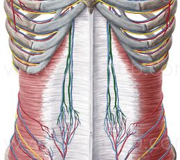
The superior epigastric arteries are important vessels of the anterior abdominal wall, being knowledge of their anatomy very important in abdominal incisions and punctures, due to the risk of injury to these vessels during procedures. However, there are controversies regarding the distribution of these vessels. The superior epigastric artery, as a branch of the internal thoracic artery, emerges in the abdomen posterior to the 7th costal cartilage, through the posterior aspect of the rectus abdominis muscle and within its sheath.
For a long time, they were not described in anatomical details. Knowledge of the topography of the superior epigastric vessels is extremely necessary for surgeons, in an attempt to reduce the risks of injuring them in abdominal incisions. The emergence of the superior epigastric arteries behind the 7th costal cartilage, within the rectus abdominis sheath.
When the superior epigastric artery is directed towards the anterior wall of the abdomen, it defines its trajectory depending on whether it is single or divided into two or more main branches.
Journey
Continues in the original direction of the internal thoracic artery , descends through the costal and sternal insertions of the diaphragm, and enters the rectus abdominis sheath, initially behind the muscle , then perforating and irrigating it, and anastomosing with the artery inferior epigastric. It emits branches that pierce the anterior wall of the sheath, supplying the muscles of the abdomen and the integument, and a small branch runs in front of the xiphoid process and anastomoses with the artery on the opposite side. It also emits small branches into the diaphragm, while from the right-sided artery, small branches extend into the falciform ligament of the liver and anastomose with the hepatic artery. . It is distributed towards the abdominal wall and the diaphragm.