A patient goes to the ophthalmology consultation when experiencing a marked symptom, such as eye pain, decreased vision, eye redness or discharge, headache , photophobia , etc.
Causes of ophthalmic conditions
Hereditary diseases
Retinitis pigmentosa
Its pathophysiology is not yet well known and, therefore, it is defined as a chronic disease, corresponding to retinal dystrophies; It is hereditary, slow and progressive, which primarily and diffusely affects the function of the photoreceptors and the pigment epithelium.
It is a chronic degenerative disease characterized by progressive loss of the visual field , night blindness and abnormal or abolished [electrorethrogram]], producing retinal atrophy, with a characteristic of pigment deposition. It affects the photoreceptors and the pigment epithelium, thus leading to blindness. It can be accompanied by other congenital affections, such as soldera, defective color vision and intellectual disorder.
- Clinical picture
Subjectively, the first clinical manifestations expressed by patients are poor night vision (nyctalopia), vision disorders in changes in lighting, as well as stumbling over objects, photophobia and vision disturbances. Progressive visual field loss is another characteristic of this disease.
The electroretinogram, initially, is subnormal. This disease can be associated with deaf-muteness, mental deficit, and hypogenitalism.
Examination of the fundus shows scattered accumulations of pigments in the periphery of the retina, with perivascular distribution, which take on the appearance of “bone spicules”, thinning of arterioles, vitreous cells and, later, pallor of the optic nerve.
- Other signs that appear are:
- Corpuscular pigmentation.
- Clusters of focal or sector pigments.
- Cystic macular edema.
- Epiretinal membrane.
- Posterior subcapsular cataract.
- Inheritance:
It can be autosomal recessive, autosomal dominant, sex-linked recessive, or simple (a single affected limb in the pedigree).
- Medical-social aspect:
Consanguinity between couples gives the medical-social aspect to the disease.
- Conduct to follow:
- Education in the community to avoid the risk of consanguinity.
- The diagnosed cases are currently undergoing medical and surgical treatment for chorioretinal revascularization, both combined, and physical-optic.
Mellitus diabetes
It is a progressive microangiopathy , it is multifactorial characterized by specific morphological changes (excavation of the disc), highlighting an acquired loss of retinal ganglion cells and their axons that affects the precapillary arterioles, capillaries and venules of the retina, it can affect major vessels. This process is also characterized by producing visual field losses and other functional changes with compromised color perception, contrast sensitivity, and mortality. It can be aggravated by pregnancy , high blood pressure , neuropathies, and anemia .
Tumors like retinobastoma
The retinoblastoma occupies 2% of all malignancies in children, has no gender predilection, generally affects only one eye, but bilateral involvement may have one of three cases. The clinical manifestations appear after 3 or 4 years of life. The disease can be inherited or due to a new genetic mutation.
Genetic load by consanguinity
This happens in diseases such as retinitis pigmentosa and sicklemia . The latter causes peripheral vessel retinopathy , sometimes extensive, posterior neovascularization, and proliferative retinopathy .
Affection of the fetus
- These occur when the pregnant woman is exposed to X-rays , as well as to the ingestion of medications:
- X-rays: It is known that exposure to pregnant women during the development of the eye can produce alterations in it.
- Ingestion of medications: The ingestion of some medications by pregnant women has teratogenic effects at the time the eye develops and even later. There are many drugs that produce toxic and degenerative effects when used.
Infectious diseases
Rubella ophthalmia
- It happens when the mother has suffered from the disease during the first three months of gestation producing cataract , microphthalmus , microcornea .
Syphilitic ophthalmia
- It is congenital or not, it can manifest as a secondary rash of the eyelids, choreoretinitis with or without interstitial keratitis is very common .
Bacterial conjunctivitis
- It is caused by any germ capable of producing acute catarrhal conjunctivitis . It can be the childhood and adult gonococcal infection . Produced by the gonococcus or another germ.
AIDS
Retinitis by fitomegalovirus is the most common intraocular infection. In patients with AIDS and the most common cause of vision loss; an increase in this condition is expected as the survival of these patients increases. It occurs when there is severe immunosuppression with a CD-4 lymphocyte count <200 cells / mm3, but it is much more frequent when the cell count is less than 100 cells.
It usually presents unilaterally at the time of diagnosis and becomes bilateral, without specific treatment.
CMV reaches the retina hematogenously and spreads in its nerve fiber layer. The patient complains of blurred vision, photophobia and visual field defects, this is depending on the degree of affectation and its location. Recurrences can occur and the evolution towards unilateral or bilateral blindness is frequent. The funduscopic image is very characteristic and consists of a single or multiple necrosis focus, extending centrifugally and encompassing vessels, with hemorrhagic involvement.
The differential diagnosis should be made with chorioretinitis caused by other herpetic viruses, mycobacteria, toxoplasmas, Candida, Pneumocystis carinii and even those caused by the human immunodeficiency virus (HIV). The diagnosis of CMV retinitis is based on funduscopic observation of the characteristic lesions. Blood, urine, and serum antibody cultures do not determine the diagnosis, given the high prevalence of seropositivity in the general adult population; furthermore, a relationship between antibody titers and CMV retinitis in AIDS patients has not been described.
- Treatment:
- Ganciclovir: 5 mg / kg, intravenous, every 12 hours, from 14 to 21 days, or foscarnet: 60 mg / kg, intravenous, every 8 hours, or 90 mg / kg, intravenous, every 12 hours, from 14 to 21 days , or ocular ganciclovir implant plus intravenous ganciclovir (the same dose recommended above), or oral ganciclovir, 1 g, 3 times a day, or cidofovir: 5 mg / kg, intravenous, weekly, for 2 weeks.
- In patients who fail monotherapy with ganciclovir or foscavir, treatment of both together will be considered, with the same doses recommended above.
Viral diseases
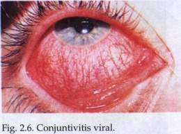
Viral conjunctivitis
Conjunctivitis caused by viral infections is quite common and generally appears in the form of an epidemic given the speed and ease of contact. There are many viruses such as enteroviruses, eye affectations causing the epidemic (che) and keratoconjunctivitis, given the speed and ease of contact; It usually affects people from the same family, schools, boarding schools, etc.
In some cases it can cause corneal scars, with significant loss of visual acuity.
Hemorrhagic conjunctivitis
Caused by enterovirus 70, it appeared in Cuba in 1981. It is highly contagious and occurs suddenly; its incubation period is 8 to 48 h. It evolves quickly and lasts 5 to 7 days.
Symptoms, as in other viral infections, are characterized by pain, photophobia, foreign body sensation, tearing, pain, photophobia. The most prominent signs are conjunctival hyperemia , pulpebral edema , chemosis, and upper vulvar conjunctival hemorrhage.
The most prominent signs are conjunctival hyperemia, eyelid edema, chemosis, and subconjunctival hemorrhages, which are located in the superior bulbar conjunctiva. Seromucosal discharge, preauricular lymphadenopathy, and conjunctival follicles are occasionally present.
This conjunctivitis usually heals spontaneously after about 7 days, therefore, patients must be isolated with strict personal hygiene measures to prevent spread. Cold boiled water compresses can be used, which help relieve symptoms and, if there is ocular discharge, the use of an antibiotic eye drop is recommended.
Trachomatous conjunctivitis
It is a chronic follicular conjunctivitis caused by Chlamydia trachomatis that is complicated by corneal opacities and ulcerations.
It is an endemic infection and a cause of blindness caused by Chlamydia trachomatis, which is a small gram-negative bacterium, an obligate intracellular parasite, that causes chronic and persistent infections.
16 Chlamydia trachomatis serotypes are known; trachoma is caused by serotypes A, B, Ba, and C, and serotypes D, E, F, G, H, I, J, K, which cause inclusion conjunctivitis.
Trachoma is a chronic follicular conjunctivitis that is complicated by corneal opacification and ulcerations. Today it is considered an immunopathological disease, caused by repeated reinfections and interrecurrent bacterial infections.
Tumors
Malignant melanoma
It is a malignant tumor that can appear on the skin, iris or choroid. It can result from the evolution of a nevus, from melanosis , as it appears primarily. It prefers to be located, initially, in the limbus, although with growth it can affect the eyelids. Its coloration is dark and varied.
- Epidemiology:
It is estimated that some 500 million people in the world are affected, many of whom are children; the disease is very common in poor areas of sub-Saharan Africa. This condition constitutes a serious health problem, since 1 to 5% of infected individuals later develop scarring that deforms the eyelids and causes ectropion with subsequent corneal damage, keratitis and blindness. In addition, it is the most commonly preventable disease, among those that cause blindness (an estimated 7 million blind people from this cause).
- Signs and symptoms:
- Photophobia.
- Blepharospasm
- Tearing
- Burning sensation and foreign body.
- Pain.
- Sometimes visual acuity disorders.
- The mucopurulent effusion is more or less abundant.
- Evolution:
The disease, due to its evolution, is divided into four periods:
- Period I (insidious). Signs and symptoms of mild conjunctivitis; thickening, edema and congestion of the conjunctiva; formation of papillae and tiny follicles in the superior tarsal conjunctiva. Vascular inflammation of the cornea may begin. Microscopic examination of the conjunctival smear shows inclusion bodies. This period lasts several weeks or months.
- Period II (acute). It is accompanied by severe inflammatory signs and symptoms (acute trachoma), profuse purulent discharge, and follicular development almost always begins in the conjunctiva of the upper eyelid. Weight gain of the eyelid causes eyelid ptosis (trachomatous ptosis). Trachomatous pannus is present in the upper part of the cornea.
- Period III (scar). It is characterized by the appearance of scar tissue, which leads to the cure of the disease. The papillae and follicles gradually disappear, but the conjunctiva does not regain its normal state. Whitish band scars persist on the tarsal conjunctiva.
- Period IV (aftermath). Represents healed trachoma. It does not always evolve favorably; relapses are frequent.
- Diagnosis:
Because most cases occur in remote areas of developing countries, the diagnosis is made clinical if two of the following appear:
- Lymphoid follicles in the superior tarsal conjunctiva.
- Lymphoid follicles along the corneal limbus.
- Scar on the conjunctival line.
- Corneal pannus.
When there is the possibility of a laboratory, this is done with the isolation of Chlamydia trachomatis in cell culture. Culture is often positive in young children with active disease; even so, the culture is only positive in 1/3 of half of the cases. Chlamydia DNA detection by polymerase chain reaction is the most sensitive test, with about 70 to 80% positivity in children with active disease. Direct immunofluorescence tests are also performed for the detection of elemental bodies (E Bis) with monoclonal antibodies, or the detection of antigens by the ELISA method.
- Treatment:
Active trachoma in children. Topical treatment with erythromycin or tetracycline, 21 to 60 days, but, as extraocular infection by Chlamydia trachomatis in the nasopharynx and gastrointestinal tract is relatively common, oral erythromycin or azithromycin, 20 mg / kg, should be indicated as a single dose in 78% of cases, it is as effective as 6 weeks of topical tetracycline or oral doxycycline, 100 mg twice a day, for 14 days, or oral tetracycline, 250 mg every 6 hours, for 14 days; Tetracycline and doxycycline should be avoided.
The application of epidemiological principles is closely linked to Ophthalmology and Optometry in relation to eye health care, to preserve individual and collective visual health. Therefore, they guide strict measures established by the Ministry of Public Health for compliance.
The prevention of communicable, contagious, non-contagious, hereditary and other conditions are determined for both the adult and the child.
- Epidemiological measures must be strictly adhered to, including:
- Isolation.
- The conjunctival exudate to know the germ and the antibiotic to use.
- The study of the environment (nursery schools, schools, work centers and barracks).
- The elimination of vectors such as flies, mosquitoes, gussases and the sanitation and cleaning of the environment.
- When there are epidemics, prophylactic treatment will be applied to healthy people in direct contact with those affected and personal and collective hygiene measures will be extreme.
- Patients will be instructed not to put dirty hands in their eyes to avoid cross-infection. It is essential to wash your hands before and after caring for patients with eye conditions.