Arteries of the masticatory apparatus. The arteries of the masticatory apparatus come from the carotid arteries. The common carotid arteries originate on the right side of the bifurcation of the bracheocephalic trunk and to the left directly from the aortic arch, both arteries ascend behind the sternoclavicular joint, vertically to the upper edge of the thyroid cartilage, where it divides into its terminal branches .
On their way through the neck , they are located in front of the transverse processes of the cervical vertebrae , being able to be compressed against the anterior tubercle of the C6 vertebra, outside the trachea , esophagus and pharynx . Behind the lobe of the thyroid gland and the homohyoid and sternocleidomasteoid muscles (satellite muscle of the artery) and inside the internal jugular vein .
The carotid inside together with the internal jugular vein, outside and behind between both the vagus nerve , are surrounded by a facial lamina called the carotid vascular sheath and the set of structures contained in the sheath constitute the neurovascular bundle of the neck.
Terminal branches of the carotid
They are the external and internal carotid. At the terminal end of the common carotid, a dilation is observed, which can be prolonged to the beginning of the internal carotid called the carotid sinus, which has abundant innervation.
The carotid glome, is the gland or paraganglion of French anatomists, it is located between the two terminal branches of the common carotid, it is made up of specific cells of the glome, connective tissue and contains a large number of vessels and nerves .
The internal carotid is used to irrigate part of the brain and the contents of the orbital cavity and small areas of the nasal mucosa .
External carotid
It extends from the common carotid fiburcation to the level of the neck of the mandibular condyle. From its origin it ascends vertically for 1 or 2 cm, then it goes slightly outwards, ascending again vertically until its termination in the thickness of the parotid gland.
Relations
In front the sternocleidomastoid muscle , the thyrolinguofacial venous trunk , behind the internal carotid, inside the pharynx , outside the digastric and stylohyoid muscles , the stylomandibular ligaments and the hyoglossal nerve . The collateral branches of the external carotid are superior thyroid, lingual, ascending pharyngeal, posterior auricular facial, occipital, and parotid.
Lingual artery
It originates above the superior thyroid, applied over the median pharyngeal contrictor, goes upwards, passes over the greater horn of the hyoid bone , enters the root of the tongue, inside the hyoglossus, goes obliquely, reaches the edge anterior of this muscle, where it divides into its terminal branches: sublingual and ranina or deep lingual.
Collateral branches
- Suprahyoid: it goes to the upper edge of the body of the hyoid bone, anastomoses with that of the opposite side and supplies the hyoid and the muscles that are inserted in it.
- Dorsal of the tongue: it goes backwards and upwards until it reaches the mucosa of the dorsal aspect of the tongue, it is distributed by the tongue, the palatoglossal arch, palatine tonsil and mucosa of the epiglottis.
Terminal branches
- Sublingual: it is directed forward through the face of the sublingual gland and above the mylohyoid muscle to which it nourishes, in addition it is distributed through the mucosa of the alveolingual sulcus, frenulum of the tongue , branch pierces the mylohyoid muscle and anastomoses with the branch submental of the facial, some twigs penetrate through the holes located around the mental spine and through the alveolar foramen to distribute in the thickness of the mandible.
- Deep lingual: it is directed forward to the tip of the tongue, situated between the lower longitudinal and the genioglossus, it is distributed through the muscles and mucosa of the tongue.
Facial artery
It emerges from the anterior aspect of the external carotid above the lingual, it goes upwards and forwards attached to the pharynx, passing through the digastric and stylohyoid muscles to the submandibular cell, contouring to the gland on its medial and superolateral, reaches the lower edge of the mandible at the level of the anteroinferior angle of the masseter and from there it goes towards the labial commissure, ascends deeply to the masogenian sulcus to the internal angle of the eye , where it anastomoses with the nasal artery of the ophthalmic, in its trajectory is very sinuous, being attached to the elevator bucacinador of the commissure and the elevators of the lip and nose.
Collateral branches on the neck
- Ascending palatine: it originates 4 or 5 millimeters from its birth, ascends through the lateral wall of the pharynx to the level of the pharyngeal orifice of the auditory tuba, is distributed by the styloglossal, style-pharyngeal muscles, walls of the pharynx, tosila palatal and mucosa of the jaws and the base of the tongue.
- Pterygoid: penetrates deep into the medial pterygoid muscle, sometimes this branch arises from the ascending palatine.
- Glandular: in variable numbers (3 or 4) it is distributed by the submandibular gland.
- Tonsillary: perforates the superior constrictor muscle of the pharynx and ends with several branches in the palatine tonsil, pharynx and root of the tongue.
- Submental: it is a thick branch, it originates from the facial at the level of the submandibular gland, it goes forward between the mylohyoid and the anterior belly of the digastricus until it reaches the chin, it is distributed by the muscles that accompany it on its way, skin and muscles of the chin and lower lip.
Facial collateral branches
- Masetheric: it originates above the lower edge of the jaw, and goes back and up on the superficial face of the masseter muscle where it ends, it also emits some twigs for the bucinator.
- Lower lip: it originates at the level of the labial commissure and goes inwards in the thickness of the lower lip.
- Upper labial: it originates at the level of the labial commissure, it goes towards anastomosing with that of the opposite side, emitting a branch for the nasal sub-septum. Both labial arteries when anastomosed with those of the opposite side, form the labial arterial ring, they are distributed by the lips.
- From the wing of the nose : it is born from the facial level of the nasal wing, it is divided into several branches for the nasal wing, back and the nasal lobe.
Terminal branch of the facial
It is the angular one which anastomoses with the nasal of the ophthalmic, emits several twigs for the skin and muscles of the nose .
Ascending pharyngeal artery
It arises from the medial aspect of the external carotid and runs up the lateral wall of the pharynx to the base of the skull. On its way it emits the branches:
- Pharyngeal: those that are distributed along the walls of the pharynx, palatine tonsil and soft palate.
- Prevertebral: aimed at this muscle group.
- Tympanic: for the eardrum .
- Posterior meningeal: which penetrates the cranial cavity through the jugular foramen, is distributed along the hypogeal nerves, vagus, superior cervical ganglion and dura mater.
Parotid arteries
They are thin branches and variable in number, they originate from the external carotid on its way through the parotid gland, in addition, small branches can also originate at the beginning of the occipital and posterior auricular.
Terminal branches of the external carotid
There are two terminal branches of the external carotid: the superficial temporal and the maxillary.
Superficial temporal artery
The direction of the external carotid continues, it leaves through the upper end of the parotid gland, between the retroarticular tubercle and the external acoustic meatus, reaching the temporal region where it divides into its terminal branches, in this region it is superficially covered by the skin .
On its way it issues the following collateral branches:
- Parotid: variable number of thin branches, emitting in the thickness of the parotid gland .
- Transverse of the face: it arises at the level of the condyle of the mandible, it goes forward following the lower edge of the zygomatic arch until it reaches the buccinator muscle, on its way it emits branches for the masseter muscle, parotid duct, facial mimic muscle and soft parts of the cheek.
- Articular: They are thin branches destined to the temporomandibular joint.
- Posterior deep temporal: it goes upwards, crosses the zygomatic arch, pierces the temporal fascia and this muscle until it is located between the muscle and the cranial wall, it is distributed through the posterior part of the muscle.
- Anterior headphones: in variable numbers they are directed backwards and are distributed by the pinna of the ear.
- Zygomatic-orbital: it is directed forward above the zygomatic arch until it reaches the lateral part of the orbicularis oculi muscle.
The terminal branches:
- Frontal: it is directed towards the region of the forehead, through which it is distributed.
- Parietal: it goes back and up to the parietal region, it is distributed in addition to the skin of this region, by that of the temporal.
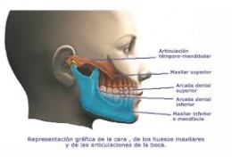
Maxillary artery
It extends from the parotid region to the pterygopalatine fossa, where it ends up continuing with the sphenopalatine.
From its origin it goes forward around the neck of the condyle of the mandible, passing through the retrocondylar eyelet, continues its journey from the inside out along the lower edge of the lateral pterygoid, attaching to the lateral aspect of the muscle, then goes up , in front and inside to the highest part of the maxillary tuberosity, then it curves inwards, penetrating the pterygopalatine fossa. As a variant in its course, the maxillary artery can be located in the deep face of the lateral pterygoid, crossing between the two fascicles of the muscle, at its anterior end from the inside out, to enter the pterygopalatine fossa by bending.
The collateral branches, according to the trajectory of the maxilla, are divided into three groups: those that originate in the mandibular, pterygoid and pterygopalatine segments.
Mandibular segment
Deep auricular: It is directed backwards and upwards and is distributed by the joint capsule of the temporomandibular joint and the inferior wall of the external acoustic meatus. Tympanic: thin branch, directed upwards, penetrates through the petrotympanic fissure, spreading through the mucosa of the eardrum.
Articular: thin branches varying in number from one to three, with an upward direction for the temporomandibular joint.
Inferior alveolar: originates close to the neck of the condyle, from its origin it goes downwards and forwards, penetrates through the mandibular foramen in the homonymous canal and runs through it in its entirety to the lower lip and buccal gingiva from premolars to incisors, and the incisor branch that continues the direction of the inferior alveolar and is distributed by the canine, incisors and bone .
In its inferior alveolar trajectory it emits the collaterals:
- Mylohyoid: it originates before the alveolar, penetrates the mandibular canal, descends through the mylohyoid groove and is distributed through the mylohyoid muscles and the anterior belly of the digastric.
- Dental: they arise from the alveolar on its way through the mandibular canal for the molars, premolars and their supporting elements.
- Bony: they are multiple branches for the mandibular bone.
Branches of the pterygoid portion of the maxilla:
- Middle meningeal: it is the thickest collateral, it is directed vertically from its beginning, it passes between the roots of the auriculotemporal nerve, passing through the spinous foramen to the cranial cavity, giving branches for the meninges, trigeminal ganglion, orbital, for the Eardrum and anastomostic branch, for the stylomastoid before penetrating the cranial cavity, it emits an interpterygoid branch from which arterioles for the pterygoid muscles arise.
- Meningeal minor or accessory: it passes into the cranial cavity through the foramen ovale, previously giving some thin branches to the lateral pterygoid and soft palate, and in the cranial cavity it is distributed through the dura near the cavernous sinus and trigeminal ganglion.
- Middle deep temporal: it arises from the maxilla at the level of the mandibular incisure, ascends outside the lateral pterygoid and enters the deep face of the temporal muscle.
- Deep anterior temporal: it is born close to the tuberosity of the maxilla in an upward direction, it is placed on the deep face of the temporal bone. The three deep temporal arteries establish anastomosis between them.
- Mesetheric: It goes outwards from its origin, crossing in front of the temporomandibular joint and through the mandibular incisure, penetrating the masseter through its deep face, in its path of articular branches. Sometimes it originates from a common trunk with the medial deep temporal.